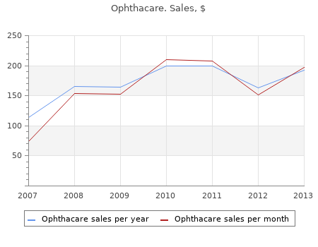
![]()

Ophthacare
By O. Giacomo. Athens State College.
Diagnosis based solely on clinical presenta- Rorabeck 10ml ophthacare sale, and Fowler, 1984). Enclosure of compartmental contents in an inelas- Any patient with clinical evidence of CECS should be tic fascial sheath considered for intracompartmental testing. Increased volume of the skeletal muscle with exer- Significant historical features include a recurrent, tion resulting from blood flow and edema exercise induced leg discomfort which increases as 3. Muscle hypertrophy as response to exercise the training persists and dissipates on cessation of 4. Dynamic monitoring is performed with the use of a An exercise challenge with detailed physical exami- slit catheter inserted prior to exertion and nation immediately after reproduction of symptoms taped/attached to the athlete’s leg for continuous will lead to a more judicious use of invasive tech- measurements. The benefit of this procedure is that niques (Glorioso and Wilckens, 2001a). There are several negative aspects of this ing both static and dynamic intramuscular pres- technique. Techniques include the needle manometer ment of catheter in the compartment during activity, (Whitesides et al, 1975), the wick catheter (Mubarak attachment of the system to the athlete, and restric- et al, 1976), slit catheter (Rorabeck et al, 1981), con- tions of the athlete’s gait as they run to reproduce tinuous infusion (Matsen et al, 1976), and a solid-state symptoms. The procedure must be performed on a transducer intracompartmental catheter (McDermott treadmill in order to continuously monitor pressure et al, 1982). Thus, the athlete cannot run outdoors on The Stryker Intracompartmental Pressure Monitor their usual training surface. In addition, only one (Stryker Corporation, Kalamazoo, Michigan) is a bat- compartment can be measured at a time. Some tery operated, hand-held, digital, fluid pressure moni- believe that with this technique, the results are tor. This device has been found to be more accurate, inconsistent and difficult to obtain and interpret versatile, convenient, and much less time consuming (Rorabeck et al, 1988; Rorabeck, Fowler, and Nott, in the clinical setting (Hutchinson and Ireland, 1999; 1988). Prior to attempting to meas- Three factors may alter the pressure measurements: ure compartment pressures, the physician should 1. Proper calibration of the monitor is essential for ensure an understanding of the anatomical structures reliable readings. The monitor must be zeroed at in each compartment so as to avoid damage to neu- the same angle that will be used to penetrate the rovascular structures.
The AP view in the infant should always be an x-ray of both hips so that the pelvic position and the horizontal situation can be evaluated generic 10 ml ophthacare visa. Guide lines for evaluating x-rays of the infant hip (Hilgen- A few guide lines will facilitate a general evaluation of reiner, Ombrédanne, acetabular angle, Shenton-Ménard); the AP view of an infant (⊡ Fig. In a dis- graphic teardrop also deforms over time if dysplasia is located hip this arc is disrupted because the femoral present. For details of the radiographic diagnosis of the hip in ▬ Acetabular roof angle = AC angle or acetabular index adolescents and adults see chapter 3. The average angle at birth Hip arthrography is suitable for evaluating the carti- is 30°, at 1 year slightly over 20° and at 3 years of age laginous sections of the hip, the ligament of head of under 20°. Although it has become less angle in infancy and early childhood, although the important since the introduction of ultrasonography, it accuracy of measurement for this angle is not very is still valuable for checking the result of a reduction and great (±5°). In particular, soft tissue obstructions in the center of the acetabulum are better evaluated by arthrography than by ultrasound. From the gluteal fold, a long needle is inserted under sterile conditions and advanced up to the hip under image-intensifier control. On the one hand it shows the whole femoral head down to the reflection of the joint capsule and, on the other, the acetabulum from the cranial labrum to the caudal acetabular rim with ⊡ Fig. The ligament of the femoral tribution in 2,294 normal and questionably pathological hips (mean, head is also shown. We can readily assess the position single and double standard deviation) of the femoral head in relation to the acetabulum and ⊡ Fig. Schematic view of the arthrographic findingsof an infant The position of the femoral head in relation to the acetabulum and with a dislocated hip: the whole femoral head down to the reflection their demarcation are readily assessed; it is also possible to establish of the joint capsule and the acetabulum from the cranial labrum to whether intra-articular soft tissue obstructions prevent the deep cen- the caudal acetabular rim with the transverse ligament, also show- tering of the femoral head ing the ligament of head of the femur (ligamentum capitis femoris). MHz transducer head for small infants and the 5 MHz It is possible to establish whether intra-articular soft head for larger infants.
Diseases buy ophthacare 10ml with visa, Pathologies, and Syndromes Defined 411 Systemic symptoms can include fever and chills, sweating, malaise, and nausea and vomiting. There may be changes in blood composition, such as an increased number of white blood cells (ie, leuko- cytes). The cortex supplied by the middle cerebral territory is most often affected (see middle cerebral artery syndrome). Occasionally, the origins of both the anterior (see anterior cerebral artery syn- drome) and middle cerebral arteries are occluded at the top of the carotid artery. Symptoms include acute abdominal cramps or steady epigastric or periumbilical abdominal pain combined with high leukocyte count. It is some- times called intestinal angina as it is the result of ath- erosclerotic plaque-induced ischemia. Intermittent back pain at the thoracolumbar junction, particu- larly with exertion, is also a common complaint. It is characterized by spontaneous bleed- ing in the absence of an identifiable precipitant and usually associated with hypertension and/or aging. IBS is referred to as nervous indigestion, functional dyspepsia, spastic colon, nervous colon, and irritable colon, but because of the absence of inflammation, it should not be confused with colitis or other inflammatory diseases of the intestinal tract. IBS is a functional disorder of motil- ity as a response to diet or stress. Kaposi’s sarcoma (KS): A malignancy of angiopoietic tissue that presents as a skin lesion. Growth of this tumor is promoted with a suppressed immune sys- tem and is an opportunistic infection associated with AIDS. Kawasaki disease: A cardiovascular pathology also known as mucocutaneous lymph node syndrome, it is an acute systemic vasculitis that can occur in any ethnic group but seems most prevalent in Asian populations. There is extensive inflammation of the arterioles, venules, and capillaries initially, then progressing to the main coronary arteries and larg- er veins. Diseases, Pathologies, and Syndromes Defined 413 Vessels develop scarring, intimal thickening, calci- fication, and formation of thrombi. This syndrome is characterized by high fever, swollen lymph nodes in the neck, rashes, irritated eyes and mucous membranes, with damage to the cardio- vascular system.
This autosomal-recessive condition is fre- tively buy cheap ophthacare 10 ml on line, and reduction by slow straightening generally quently found in Arab countries. The feet also usually require surgery to correct the pronounced equinus deformity. In addition to This inherited disorder is characterized by a flat face, fixed scoliosis, an atlantoaxial instability can also develop, bulging forehead, hypertelorism and multiple congenital requiring early occipitocervical fusion. Both autosomal-dominant and autosomal-recessive Mucopolysaccharidoses inheritance patterns have been observed. The gene locus > Definition in the more common dominant variant is 3p21. It The mucopolysaccharidoses form a group of conditions appears to involve a generalized mesenchymal defect that involving defective lysosomes. The disease is very involved in mucopolysaccharide metabolism, and their rare and the literature only contains isolated cases. There failure can lead to the storage of mucopolysaccharide is a striking accumulation in La Réunion, where 38 cases components. Classification, occurrence, etiology The tarsal bones often show multiple ossification ⊡ Table 4. A tracheomalacia in infancy and early child- doses in six types, based on the enzyme defect and list- hood can cause major problems. The authors of a 30-year study in by malformations that lead to kyphosis or scoliosis Great Britain calculated a prevalence for mucopolysac- (⊡ Fig. Historical background ▬ Differential diagnosis: Larsen syndrome can be con- Type I mucopolysaccharidosis was first described by Gertrude Hurler in the year 1919. The term »gargoylism« was coined by Ellis, Sheldon fused with arthrogryposis multiplex congenita, in and Capon, and refers to gargoyles, those grotesque figures on which the joints can also be severely deformed or Gothic cathedrals that spit out the rainwater. A pronounced stiffness is generally present was published by Hunter in 1917, while type III (Sanfilippo syn- in arthrogryposis however, which is not the case with drome) was first mentioned in 1961 by Harris and described in 1963 by Sanfilippo [33, 104]. Since significant ligament laxity Morquio and Brailsford in 1929.
Athens State College.